


L'endophthalmitis is a rare but serious form of inflammation of the eyeballdue to a infection of the intraocular cavity which, if not treated adequately and in a timely manner, can lead to irreversible loss of vision.
Classification
Based on the route of infection transmission, endophthalmitis can be distinguished into exogenous or endogenous.
Exogenous endophthalmitis
It is defined as exogenous endophthalmitis direct infection of the eye by microorganisms that enter the intraocular cavity through trauma or eye surgery.
According to some studies, exogenous infection occurs in 11% of open-eye injuries and in approximately 0.1% of eyes undergoing intraocular surgery.
Postoperative infectious endophthalmitis
L'postoperative infectious endophthalmitis is, in fact, one of the most feared complications of eye surgery. Furthermore, exogenous infection can in turn be distinguished into acute or chronic.
L'acute exogenous infection occurs a few days after the event and is extremely painful.
On the contrary, thechronic exogenous infection can develop months or even years after surgery (the average is about 9 months), is less common than acute and has a better prognosis due to the less severe nature of the inflammation.
Endogenous endophthalmitis
L'endogenous endophthalmitis arises when the infection of the eye is indirect, i.e. by micro-organisms that have spread through the bloodstream but originate from a distant infected site, e.g. the heart (endocarditis).
Typical micro-organisms that cause endogenous infection are bacteria such as Staphylococcus aureus, Streptococcus pneumoniae, Streptococcus viridans, Escherichia coli and fungi such as Candida. Endogenous infection is rarer than exogenous: an estimated incidence of 5 per 10,000 hospitalised patients.
Usually one occurs in debilitated individuals and who are unable to activate an immune response against infection, such as drug users, chemotherapy patients and organ transplant recipients taking immunosuppressive drugs.
Other risk factors include AIDS, poorly controlled diabetes mellitus and long-term steroid use.
Symptoms of endophthalmitis
In most cases, irrespective of its origin, endophthalmitis mainly presents with eye pain and reduced or blurred vision, is accompanied by photophobia, swelling of the eyelids, chemosis, redness.
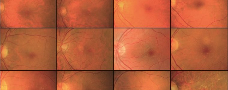
In some cases, pus may be present in the anterior chamber of the eye (the area between the iris and the lens), vitreitis (inflammation of the vitreous body) and retinal periphlebitis (inflammation of the blood vessels of the retina).
Diagnosis
In the case of suspected endophthalmitis, an early diagnosis is essential.
Usually, the diagnosis of post-operative endophthalmitis is immediate as soon as one visits the ophthalmologist. Already at the slit lamp it is possible to realise whether an infection is the cause of the patient's symptoms.
I local symptoms and signs which are of varying intensity are ocular and periocular pain, photophobia, severe visual impairment to the point of light perception; this may be due to either opacification of the ocular dioptric media (corneal oedema, fibrin and deposits on the IOL, vitreal turbidity) or anatomical damage to retinal structures (abscesses, necrosis, oedema).
The patient may report also systemic symptoms and signs as headache, fever, vomiting, physical prostration. Objectively the patient may present with: perikeratic injection, eyelid oedema of varying intensity, chemosis, conjunctival hyperemia and secretions, corneal oedema and endothelial precipitates, cellular tyndall and hypopion in the anterior chamber, reduction or disappearance of the pupillary reflex, fibrin in the pupillary field, fibrin deposits on the IOL, retinal haemorrhages, vitreous cloudiness and thickening with unexplored fundus, chorioretinal foci, retinal abscesses and/or necrosis, ocular hypertone or hypotone, decreased response to cycloplegics, Seidel's + test (in post-surgical forms and secondary to perforating trauma) for de-escalation of wound margins.
Endophthalmitis may evolve into panophthalmitis, involving all ocular structures and in some rare cases the inflammatory process may also involve the parabulary structures.
Ultrasound examination and microbiological tests
In the presence of pronounced opacity of the dioptric media, an ultrasound examination (A-B- scan, UBM) can be used to assess the presence of inflammatory material in the posterior segment, the severity and extent of inflammation, the degree of involvement of the endoocular structures, the possible presence of cortical debris in the vitreous chamber and the presence of complications (retinal detachment, choroid detachment). Acute postoperative infectious endophthalmitis initially remains a clinical diagnosis and is considered presumptive until established by Gram stain, culture or PCR. Although endophthalmitis must be confirmed by microbiological laboratory analysis, it must be considered an ophthalmic emergency. It is therefore advisable to intervene as soon as possible after clinical diagnosis because the bacteria replicate exponentially and their toxic by-products, with the associated inflammatory state, are capable of destroying visual capacity.
Endophthalmitis: treatment
Depending on the severity of the infection, treatment includes administration of antibiotic, anti-fungal and/or antiviral drugs topically, orally, intravenously or by direct injection into the eye. If the infection is severe, a surgery vitrectomy in which the vitreous body (gelatinous liquid contained in the eyeball cavity) is removed and replaced with a similar medium (vitreous substitute).
See also
Endophthalmitis: decreasing rates - Oculista Italiano
–Endophthalmitis: Pathogenesis, clinical presentation, management, and perspectives. Clinical Ophthalmology. 2010