


The glaucoma is a chronic progressive, sight-threatening disease that requires the use of topical drugs to lower intraocular pressure (IOP), to be administered for decades and, in many cases, for life.
Pathology
Glaucoma is a chronic degenerative disease affecting the optic nerve, characterised by damage to its nerve fibres and consequent visual field damage. If left untreated, the progressive reduction of the visual field can lead to blindness.
Prevalence
Glaucoma is a major social problem: it is the second leading cause of blindness in the world and affects about 60 million people, rendering more than 8 million of them blind. Moreover, about 50% of glaucoma sufferers are unaware of it.
Early Diagnosis
It is a sneaky disease as one often notices the disease when visual changes are already very advanced, whereas previously one did not experience any symptoms. However, if diagnosed early and properly treated, it can be effectively kept under control, allowing good vision throughout life.
Risk factors
Numerous risk factors correlate with the onset of the disease. Among these, the main ones are:
- elevated intraocular pressure (IOP),
- advanced age,
- familiarity.
Intraocular pressure
The IOP value is determined by a fluid circulating within the eye, the aqueous humour. In a healthy eye, the ratio of aqueous humour produced to aqueous humour excreted is such that a constant intraocular pressure is maintained, usually between 11 and 20 mmHg.
In the presence of glaucoma, on the other hand, this relationship is altered due to a reduction in the elimination of aqueous humour that occurs at the level of the trabecular meshwork (structure that allows this fluid to escape from the eye).
Classification
Glaucoma can be classified in several ways:
- Depending on the aetiology, glaucoma can be primary, when it occurs in the absence of other ocular or systemic pathologies, or secondary, when associated with pre-existing pathologies;
- Based on the altered outflow of the aqueous humour, we distinguish between open-angle glaucoma, due to increased resistance to outflow at the level of the trabecular meshwork, and angle-closure glaucoma, where there are anatomical problems that prevent the aqueous humour from reaching the trabecular meshwork;
- Based on the value of the main risk factor, IOP, there is high-pressure glaucoma and normal-pressure glaucoma.
Added to these are congenital or acquired glaucoma, if IOP is elevated from birth, and infantile glaucoma if it occurs during the first years of life.
Symptoms
The increase in intraocular pressure and the resulting damage to the optic nerve are not perceptible and, therefore, the main symptom is a progressive narrowing of the visual field.
Unfortunately, in the early stages, it is not possible to realise this limitation without an eye examination.
Diagnosis
There are several tests that can be used to rule out or diagnose the presence of glaucoma:
1. the tonometry, which is used to measure intraocular pressure;
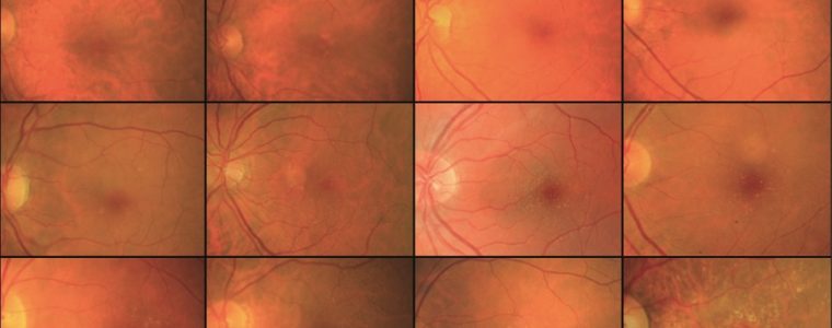
2. l'ophthalmoscopywhich allows an objective examination of the optic nerve;
3. the perimeter (or visual field examination), which allows an assessment of overall visual function;
4. the pachymetrywhich assesses corneal thickness;
5. OCT, which evaluates the retinal nerve fibre layer.
Apart from measuring IOP, some of these tests are necessary to monitor the evolution of the disease.
Treatment
Glaucoma therapy involves two types of approach, pharmacological and surgical.
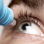
Pharmacological treatment is the first choice and uses anti-glaucomatous drugs aimed at reducing the main risk factor for glaucoma, intraocular pressure.
These drugs are taken as chronic therapy, i.e. administered regularly and consistently throughout life.
Full adherence to therapeutic prescriptions is essential for the treatment to produce its effects.
In the event that pharmacological therapy alone fails to reach a certain target pressure, parasurgical therapy (laser treatments) or, alternatively, surgical treatment can be used.
The most common surgery is called trabeculectomy and involves the creation of an artificial aqueous humour outflow channel.
Topical drugs and side effects
Topical glaucoma drugs are associated with a significant incidence of ocular surface disease (OSD).
Importantly, almost half of glaucoma patients worldwide receive more than one drug to lower IOP, with gradual implementation of multiple topical agents to reduce and achieve the desired target pressure.
The different treatment options
Today, a multitude of treatment options are available, which can be administered as fixed combinations or simultaneously.
However, active compounds, individually or in combination, their excipients, and in particular preservatives, interact in complex ways with the ocular surface.
Managing and optimising ocular surface health is, therefore, a major challenge in the management of glaucoma.
Glaucoma treatment and OSD: the correlation
Many studies Observational studies have shown that the prevalence of glaucoma therapy-related OSD is much higher than that found in the general population, with as many as 45-60% of patients using preservative eye drops being clinically affected by ocular surface disease.
The effects on the ocular surface
Long-term use of topical IOP-lowering drugs can induce ocular discomfort, tear film instability, Meibomian gland dysfunction, conjunctival inflammation, subconjunctival fibrosis, epithelial damage and allergic blepharitis.
Furthermore, it is plausible that the ocular surface is damaged at various levels by subtle subclinical inflammation, observed in the 80% of pharmacologically treated glaucoma patients, which can cause serious consequences, up to devastating diseases such as toxic pseudopemphigoid.
In addition, OSD may reduce long-term tolerance and adherence to topical therapy, due to a wide range of adverse events occurring within a few months of starting treatment.
Benzalkonium chloride (BAK) toxicity
A large number of clinical and experimental studies have documented a strong correlation between signs and symptoms of OSD with the number of glaucoma medications used and, in particular, with cumulative exposure to benzalkonium chloride (BAK). In fact, most dispensers of standard multi-use eye drops contain BAK as a preservative.
Several evidences suggest that on the ocular surface, BAK causes tear film instability, loss of calico cells, conjunctival squamous metaplasia, cell apoptosis, disruption of the corneal epithelial barrier, corneal nerve damage and potential damage to deeper ocular tissues, where it gradually accumulates. These pathological changes to the ocular surface may also be associated with subsequent conjunctival fibrosis, and thus increase the risk of filtering surgery failure, especially in those patients treated with multiple BAK drugs stored for many years.
Preservative-free drugs for the treatment of glaucoma: a possible solution
Growing data suggest, therefore, that prevention or amelioration of OSD can be achieved by eliminating BAK exposure through the use of preservative-free glaucoma eye drops (PF).
Preservative-free (PF) drugs are a viable treatment strategy in the ongoing management of glaucoma. By removing the toxicity of preservatives, PF formulations provide tangible clinical benefits. In addition, they improve tolerability and adherence, leading to a positive impact in long-term intraocular pressure (IOP) control.
For example, to date, for most patients with glaucoma or ocular hypertension, the initial topical eye drop of choice is a prostaglandin analogue (PGA), due to its superior 24-hour efficacy and satisfactory tolerability profile. Undoubtedly, prostaglandins, enzyme products of essential fatty acids, show an excellent overall safety profile, however they exhibit a number of ocular side effects, such as conjunctival hyperemia, iris hyperpigmentation, periocular skin darkening, hypertrichosis and OSD. In contrast, patients treated with PGA without conservatives have been shown to develop fewer adverse effects. The same was observed for prostaglandin/timolol combinations without preservatives.
Studies on pilocarpine, beta-blockers, CAI and brimonidine and on combinations, albeit in smaller numbers, showed similar results regarding safety and tolerability of the preservative-free versions.
Overall, therefore, the switch from preservative to PF drugs offers the significant advantage of reducing the side effects caused by the preservative agent. The improved tolerability is observed in clinical signs, as well as in the improvement of patients' quality of life. The reduction in adverse effects also improves adherence to therapy and supports physicians in the management of their glaucoma patients.