
In glaucoma, every hour that passes reduces the chances of successful therapy.
What are the clinical signs
If your dog or cat shows any of the clinical signs listed below, the first thing to do is to have it examined immediately (or as soon as possible) by your vet, preferably an ophthalmologist.
- Rednesscaused by congestion of the venous vessels of the sclera (the 'white' of the eye around the cornea will appear congested)
- Corneal opacity. Increased intraocular pressure can damage the endothelium, the innermost layer of the cornea (the transparent 'glass' at the front of the eye). As a consequence, oedema can occur, which will cause the cornea to appear bluish, the famous 'glaucous' colour from which the disease derives its name.
- Pain. In dogs and cats it may not be immediately recognisable, but it can be very intense indeed. Human glaucoma patients report sometimes unbearable fierce headaches. Depending on the basic temperament, our animal patient may be extremely depressed or particularly reactive/aggressive, showing particular impatience when touched around the eye. Let us not forget that dogs and cats cannot talk to each other, so let us never underestimate sudden changes in the behaviour of our pet. And when, as we will discuss later, the clinical situation is extreme and requires removal of the eye, let us never forget that owners often report an extreme improvement in the patient's quality of life following surgery to remove the glaucomatous eye (enucleation).
- Visual impairment or sudden blindness. The marked increase in intraocular pressure causes significant suffering of the retina and optic nerve, which reduces visual capacity to the point of complete blindness. Contrary to what one may believe, referring to humans, it is not easy to notice blindness (especially if only of one eye) in our pets. Especially in the domestic environment, dogs and cats are often able to orient themselves very well without the aid of sight. We may perhaps only become aware of the reduced visual ability by changing the position of objects/movements along their usual route,
- Increased eye volume. The strong increase in pressure can cause dilation of the collagen fibres of the sclera and cornea, leading to an increase in the size of the eye (buftalmos). This sign is not early and usually manifests itself in the course of chronic (i.e. very advanced) glaucoma or in very young subjects.
- Pupil dilation. In the early stages it may be moderately dilated, and still respond to light, albeit incompletely. As the disease progresses or in acute cases with very high pressure, the pupil is completely dilated and does not respond to light. The owner will have the impression that he or she can no longer see the colour of the eye or that the eye is 'blacker'.
- Lens dislocation. The crystalline lens may move from its anatomical position (behind the iris, immediately in front of the vitreous) due to the increase in volume of the eye, which causes the fibres holding it in place to break. The opposite can also happen, i.e. the dislocated lens (due to genetic predisposition, trauma, chronic uveitis) can be the cause of glaucoma.
Diagnosis of glaucoma
The instrument needed to measure intraocular pressure, the electronic tonometer (either magnetic rebound or applanation), is generally not available in basic veterinary facilities, which is why a specialist ophthalmological examination is recommended.
The eye examination
A comprehensive eye examination will allow not only the diagnosis to be made, but also the prognosis to be assessed.
Specific examinations
In some cases, it may be necessary to carry out special investigations, which we explain in detail below
Gonioscopy
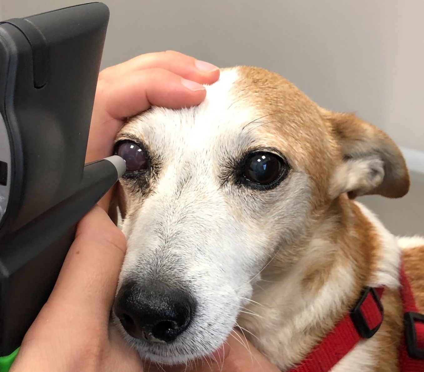 Gonioscopy involves the use of a special lens that, when applied to the cornea, allows visualisation of the iridocorneal angle, the area through which aqueous humour drains. It is mainly performed in cases where primary glaucoma is suspected, based on clinical history and race. Generally, only local anaesthesia is sufficient, so that the gonioscopy lens can be placed on the cornea without bothering the patient too much.
Ocular ultrasound
Ocular ultrasound can be useful in cases where the eye is already opaque and it is not possible to visualise intraocular structures. Even in this case, local anaesthesia is usually sufficient to allow the probe to be placed directly on the cornea or eyelids that are kept closed.
Prognosis
Unfortunately, glaucoma is an irreversible disease, unless the underlying cause can be removed, such as in the case of dislocation of the crystalline lens, where the lens can be surgically removed. This means that medical therapy will have to be adopted for the rest of the patient's life.
Therapy
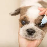
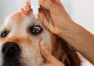
Therapy generally consists of a combination of medication (mostly eye drops) and regular check-ups at the veterinary ophthalmologist.
In selected cases, a surgical approach is also available. Unfortunately, despite therapeutic efforts, it is often difficult to keep glaucoma under control and medical therapy may no longer be effective within a variable timeframe.
If the pressure is permanently high despite therapy, intraocular pain is also considerable, and in the case of blindness, enucleation is the best indication for treating chronic pain.
Medical therapy
 The treatment of glaucoma can be medical, surgical or associated. Medical therapy is undoubtedly the first approach and must be immediate. The main aim is to lower the intraocular pressure to levels that the eye can tolerate, treat the underlying cause (if it exists and is possible), reduce the risk of blindness as much as possible, trying to preserve as much vision as possible and, last but not least, control pain.
Therapy must always be set up under the direct and strict control of the veterinary doctor, and do-it-yourself therapies and advice from 'who knows why his dog/cat too...' should be absolutely avoided. Each individual is a separate case and must be followed by the doctor.
The medical therapy mainly consists of the administration of eye drops that reduce the production of aqueous humour or increase its drainage via the unconventional route.
Antiglaucomatosis drugs
Osmotic diuretics
Mannitol is used ONLY in emergencies in the treatment of acute glaucoma. It is administered intravenously and requires specialist attention.
Synthetic prostaglandin analogues
They increase drainage through the unconventional route, and are therefore generally quite effective. Administered once or twice a day, Latanoprost, Travoprost and the like can be used long-term
Carbonic anhydrase inhibitors
They reduce aqueous humour production, thus reducing intraocular pressure. These include Dorzolamine and Brinzolamide. Oral drugs do not show superior efficacy and are a potential cause of systemic side effects.
β-blockers
By reducing blood flow to the ciliary bodies, they reduce the production of aqueous humour. Not are among the most widely used in veterinary ophthalmology, but may have synergistic effects with other topical therapies. Especially in small animals they may have side effects, particularly if concomitant pulmonary or cardiac diseases are present. These include Timolol and Betaxolol.
We look forward to the third instalment to consider cases where medical therapy fails to bring the condition under control.
We will see together what is best to do and how best to manage our sick animal.
Daniele Santillo
Dr. Daniele Santillo
Med Vet, CertVOphthal, MRCVS
RCVS Advanced Practitioner in Veterinary Ophthalmology
Arezzo - Santillo short CV
In this section:
- Focus on: veterinary ophthalmology
- The opaque eye: not only cataracts...
- Opaque eye: could be glaucoma?
- Glaucoma: when medical therapy does not work...